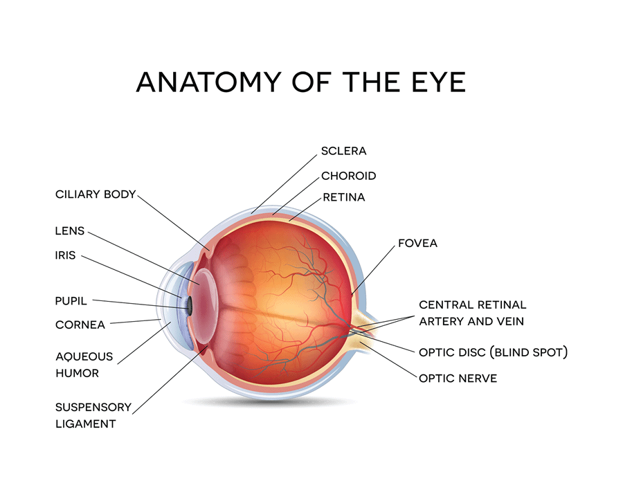
Optic Disc On Eye Diagram . It is where the retina and optic nerve connect. the optic disc is an elevation on the medial aspect of the retina where the sensory fibers and retinal vessels pass. the structure around the optic nerve where it enters the back of the eye. the optic disc, sometimes called the optic nerve head, is a round section at the back of the eye. the optic disc represents the beginning of the optic nerve (second cranial nerve) and is the point where the axons of retinal ganglion cells come together. the optic disc is where the axons of retinal ganglion cells join together and mark the beginning of the optic nerve (second cranial nerve). Read an overview of general eye anatomy to learn how the parts of the.
from www.feelgoodcontacts.ie
Read an overview of general eye anatomy to learn how the parts of the. the optic disc is an elevation on the medial aspect of the retina where the sensory fibers and retinal vessels pass. It is where the retina and optic nerve connect. the optic disc, sometimes called the optic nerve head, is a round section at the back of the eye. the optic disc is where the axons of retinal ganglion cells join together and mark the beginning of the optic nerve (second cranial nerve). the optic disc represents the beginning of the optic nerve (second cranial nerve) and is the point where the axons of retinal ganglion cells come together. the structure around the optic nerve where it enters the back of the eye.
Anatomy of human eyehow the human eye works Feel Good Contacts
Optic Disc On Eye Diagram the optic disc is where the axons of retinal ganglion cells join together and mark the beginning of the optic nerve (second cranial nerve). Read an overview of general eye anatomy to learn how the parts of the. the optic disc is an elevation on the medial aspect of the retina where the sensory fibers and retinal vessels pass. the optic disc represents the beginning of the optic nerve (second cranial nerve) and is the point where the axons of retinal ganglion cells come together. the structure around the optic nerve where it enters the back of the eye. the optic disc is where the axons of retinal ganglion cells join together and mark the beginning of the optic nerve (second cranial nerve). It is where the retina and optic nerve connect. the optic disc, sometimes called the optic nerve head, is a round section at the back of the eye.
From www.researchgate.net
Fundus photo of the left eye Optic disc is faded; retinal vascular... Download Scientific Diagram Optic Disc On Eye Diagram the optic disc represents the beginning of the optic nerve (second cranial nerve) and is the point where the axons of retinal ganglion cells come together. It is where the retina and optic nerve connect. the optic disc, sometimes called the optic nerve head, is a round section at the back of the eye. the optic disc. Optic Disc On Eye Diagram.
From dxovvkact.blob.core.windows.net
Optic Disc Definition Eye at Daniel Binder blog Optic Disc On Eye Diagram the optic disc is an elevation on the medial aspect of the retina where the sensory fibers and retinal vessels pass. the structure around the optic nerve where it enters the back of the eye. Read an overview of general eye anatomy to learn how the parts of the. the optic disc represents the beginning of the. Optic Disc On Eye Diagram.
From geekymedics.com
The Optic Nerve (CN II) Cranial Nerve II Geeky Medics Optic Disc On Eye Diagram the optic disc, sometimes called the optic nerve head, is a round section at the back of the eye. the optic disc is where the axons of retinal ganglion cells join together and mark the beginning of the optic nerve (second cranial nerve). the structure around the optic nerve where it enters the back of the eye.. Optic Disc On Eye Diagram.
From dxovvkact.blob.core.windows.net
Optic Disc Definition Eye at Daniel Binder blog Optic Disc On Eye Diagram the optic disc represents the beginning of the optic nerve (second cranial nerve) and is the point where the axons of retinal ganglion cells come together. the optic disc is where the axons of retinal ganglion cells join together and mark the beginning of the optic nerve (second cranial nerve). the optic disc, sometimes called the optic. Optic Disc On Eye Diagram.
From pressbooks.bccampus.ca
11.1 Physics of the Eye and the Lens Equation Douglas College Physics 1207 Optic Disc On Eye Diagram the optic disc is where the axons of retinal ganglion cells join together and mark the beginning of the optic nerve (second cranial nerve). It is where the retina and optic nerve connect. the optic disc, sometimes called the optic nerve head, is a round section at the back of the eye. the optic disc represents the. Optic Disc On Eye Diagram.
From www.wisegeek.com
What is the Optic Disc? (with pictures) Optic Disc On Eye Diagram Read an overview of general eye anatomy to learn how the parts of the. the optic disc is where the axons of retinal ganglion cells join together and mark the beginning of the optic nerve (second cranial nerve). It is where the retina and optic nerve connect. the optic disc represents the beginning of the optic nerve (second. Optic Disc On Eye Diagram.
From www.dreamstime.com
Normal Eye Retina, Illustration Stock Illustration Illustration of retinal, investigation Optic Disc On Eye Diagram the optic disc, sometimes called the optic nerve head, is a round section at the back of the eye. Read an overview of general eye anatomy to learn how the parts of the. the structure around the optic nerve where it enters the back of the eye. the optic disc is where the axons of retinal ganglion. Optic Disc On Eye Diagram.
From www.mitchmedical.us
Normal Optic Disc Physical Diagnosis Mitch Medical Optic Disc On Eye Diagram the optic disc is an elevation on the medial aspect of the retina where the sensory fibers and retinal vessels pass. It is where the retina and optic nerve connect. the optic disc, sometimes called the optic nerve head, is a round section at the back of the eye. the optic disc is where the axons of. Optic Disc On Eye Diagram.
From exohqvonz.blob.core.windows.net
Eye Optics Diagram at Florence Gaston blog Optic Disc On Eye Diagram Read an overview of general eye anatomy to learn how the parts of the. the optic disc, sometimes called the optic nerve head, is a round section at the back of the eye. the optic disc is an elevation on the medial aspect of the retina where the sensory fibers and retinal vessels pass. the optic disc. Optic Disc On Eye Diagram.
From www.researchgate.net
Normal appearance of optic disc in the right eye Download Scientific Diagram Optic Disc On Eye Diagram the optic disc, sometimes called the optic nerve head, is a round section at the back of the eye. the optic disc is an elevation on the medial aspect of the retina where the sensory fibers and retinal vessels pass. the optic disc is where the axons of retinal ganglion cells join together and mark the beginning. Optic Disc On Eye Diagram.
From www.pinterest.co.uk
Roles of Optic Disc Diagnosis Optometry, Eye facts, Optometry education Optic Disc On Eye Diagram the structure around the optic nerve where it enters the back of the eye. the optic disc is an elevation on the medial aspect of the retina where the sensory fibers and retinal vessels pass. the optic disc is where the axons of retinal ganglion cells join together and mark the beginning of the optic nerve (second. Optic Disc On Eye Diagram.
From www.researchgate.net
Anatomical structure of optic disc. Download Scientific Diagram Optic Disc On Eye Diagram the optic disc is where the axons of retinal ganglion cells join together and mark the beginning of the optic nerve (second cranial nerve). It is where the retina and optic nerve connect. the optic disc represents the beginning of the optic nerve (second cranial nerve) and is the point where the axons of retinal ganglion cells come. Optic Disc On Eye Diagram.
From www.shutterstock.com
2,406 Optic Disc Images, Stock Photos & Vectors Shutterstock Optic Disc On Eye Diagram the optic disc represents the beginning of the optic nerve (second cranial nerve) and is the point where the axons of retinal ganglion cells come together. Read an overview of general eye anatomy to learn how the parts of the. the structure around the optic nerve where it enters the back of the eye. It is where the. Optic Disc On Eye Diagram.
From www.knowyourbody.net
Optic Nerve Definition, Function, Anatomy and FAQs Optic Disc On Eye Diagram the optic disc is an elevation on the medial aspect of the retina where the sensory fibers and retinal vessels pass. the optic disc is where the axons of retinal ganglion cells join together and mark the beginning of the optic nerve (second cranial nerve). the optic disc represents the beginning of the optic nerve (second cranial. Optic Disc On Eye Diagram.
From gene.vision
Optic nerve Gene Vision Optic Disc On Eye Diagram the optic disc is where the axons of retinal ganglion cells join together and mark the beginning of the optic nerve (second cranial nerve). the optic disc is an elevation on the medial aspect of the retina where the sensory fibers and retinal vessels pass. Read an overview of general eye anatomy to learn how the parts of. Optic Disc On Eye Diagram.
From www.researchgate.net
A CASE OF OPTIC DISC; RIGHT EYE Download Scientific Diagram Optic Disc On Eye Diagram the optic disc, sometimes called the optic nerve head, is a round section at the back of the eye. the structure around the optic nerve where it enters the back of the eye. the optic disc is where the axons of retinal ganglion cells join together and mark the beginning of the optic nerve (second cranial nerve).. Optic Disc On Eye Diagram.
From anatomicaljustice.com
Cupped Optic Disc of the Right Eye No Text Optic Disc On Eye Diagram It is where the retina and optic nerve connect. Read an overview of general eye anatomy to learn how the parts of the. the optic disc is an elevation on the medial aspect of the retina where the sensory fibers and retinal vessels pass. the optic disc represents the beginning of the optic nerve (second cranial nerve) and. Optic Disc On Eye Diagram.
From commontastebuds.com
Eye Anatomy Understand how your eyes work to produce one of the most important senses, vision. Optic Disc On Eye Diagram Read an overview of general eye anatomy to learn how the parts of the. the optic disc is an elevation on the medial aspect of the retina where the sensory fibers and retinal vessels pass. the optic disc is where the axons of retinal ganglion cells join together and mark the beginning of the optic nerve (second cranial. Optic Disc On Eye Diagram.